


The following information was adapted from a July, 1990 article entitled Mosquito-Borne Dog Heartworm Disease by Dr. Jai K. Nayar, Florida Medical Entomology Laboratory, IFAS-University of Florida, 200 9th Street, Southeast, Vero Beach, FL 32962. Phone: (561) 778-7200, Fax: (561) 778-7205. Additional information also may be obtained from Dr. Charlie Morris, Extension Entomologist, Florida Medical Entomology Laboratory, IFAS-University of Florida, 200 9th Street, Southeast, Vero Beach, FL 32962, Phone and Fax (561) 778-7204.
Each year thousands of dogs become disabled or die from lung, heart or circulatory problems caused by the heartworm disease. Heartworm disease in dogs and related canines is caused by a filarial nematode (thread-like round worm), Dirofilaria immitis. It is a major problem that promises to become more serious with time. The adult inhabits the right ventricle and pulmonary arteries, and because of its location in the heart, it is commonly called "the dog heartworm." The parasite can only be transmitted from one dog to the other by some vector mosquito species.
 DISTRIBUTION OF HEARTWORM DISEASE:
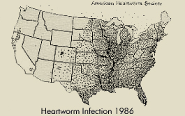
Heartworm is distributed worldwide in most tropical and subtropical regions, with increasing frequency in temperate climates. Until the late sixties, the disease was restricted to southern and eastern coastal regions of the United States. Now, however, cases have been reported in almost every state and in several provinces of Canada as shown here.
For most of North America, the danger of infection is greatest during the summer when temperatures then are favorable for mosquitoes. In the southern U.S., especially the Gulf Coast and Florida, where mosquitoes are present year-round, the threat of heartworm disease is constant.
The complete development of the nematode parasite requires two hosts: the dog and the mosquito. The problem starts when a mosquito draws blood from an infected dog. In the dog, sexually mature adult nematodes are 8 to 14 inches long and live in the heart. Once a dog is infected, it is infected for life. The sexually mature nematodes deposit tiny immature worms called microfilariae, which circulate in the blood stream. Microfilariae are less than 1/800 of an inch long. They do not develop further in the dog, but they can survive in circulation for up to seven years. They must be ingested by a mosquito before they can progress in their development. Numbers of microfilariae in blood are generally higher during the day than at night. Optimum numbers of microfilariae in the peripheral circulation coincide with times of peak feeding activity by the mosquito vector. Numbers of microfilariae may be higher in the summer when mosquitoes are abundant.
Development of heartworm in the vector starts when microfilariae are ingested by the female mosquito during blood feeding on an infected dog. Microfilariae leave the midgut of the mosquito soon after ingestion and migrate into the Malpighian tubules cells (the mosquito kidney). During the next four days the parasite becomes immobile, shortens and thickens, and gives rise to the so-called "sausage form" larva.
This larval form is followed by the first stage larva and the first molt occurs in the Malpighian tubule cells at eight days. During the second larval stage, formation of internal organs takes place. The second molt occurs at twelve days. Third-stage larvae resemble miniature adults. During the next two to three days, they increase in length, break out of the Malpighian tubules, migrate through the body to the head, and accumulate in the mouthparts. These third-stage larvae are now called "infective larvae" (Fig. 2).
Thus, in two to three weeks, a microfilaria transforms into an infective larva. This infective larva cannot develop further in the mosquito. Further development can only take place in a dog. Infective larvae are found primarily in the proboscis, or mouth parts. As the infective mosquito feeds on a dog, the infective larvae emerge from the timp of the proboscis and on to the skin of the animal. A drop of mosquito blood protects the larvae from drying prior to their entry into a host. The infective larvae penetrate the skin through the puncture wound that remains after the mosquito withdraws her mouthparts.
After penetrating the skin, the larvae stay close to the site and grow very little during the next few days. The molt from third-to-fourth stage larvae occurs six to ten days after infection. Fourth-stage larvae migrate through subcutaneous tissue and muscle toward the upper abdomen and thoracic cavity. Fourth-stage larvae grow to about 1/10 of an inch in length during the next 40 to 60 days and then molt to the fifth and final larval stage, or young adults. The young adults penetrate veins to get into the blood stream and eventually, after 70 to 90 days in the dog, reach the heart. For unknown reasons, the percentage of infective third-stage larvae that reach maturity vary in different breeds of dogs. Upon reaching the heart, the young adults continue to grow. Up to now there has been no evidence of disease in the dog. It is only after adult worms mate and start to deposit tiny motile microfilariae that circulate in the blood that disease becomes apparent. Microfilariae appear in the blood about 200 days after infection.
Visible signs of heartworm disease may not appear until a full year after infection. In fact, the disease may be well advanced before the dog shows any symptoms. Dogs with typical heartworm disease fatigue easily, cough, and appear rough and un-thriving. Blood and worms from ruptured vessels may be coughed up. Blockage of major blood vessels can cause the animal to collapse suddenly and die within a few days. Dogs with 50 to 100 mature worms exhibit moderate to severe heartworm disease. Dogs with 10 to 25 worms that receive little exercise may never show signs of heartworm disease, and one may not be able to find microfilaria in the blood. Heartworm infection without detectable microfilaremia is called occult dirofilariasis.
Diagnosis of dog heartworm disease is done by drawing a blood sample and looking for microfilaria using a microscope. These tests are reliable only 80% of the time. A more reliable method is to take x-rays. When heartworm disease in confirmed, a treatment program is set up to remove both adult worms and microfilariae.
Heartworm disease in cats is less frequent than in dogs. Cats are susceptible, but appear to be poorer hosts than the dog. The most prominent clinical signs include coughing, dyspnea, vomiting, lethargy, and anorexia. Acute collapse and death can occur. Because less than 20% of infected cats have microfilaria in the blood, diagnosis is best confirmed by x-rays.
Heartworm is also an occasional parasite of humans. The parasite is usually found in the lung (pulmonary dirofilariasis), less often in the heart. Although the worm forms a "coin lesion" in the lung which may be confused with other diseases on x-rays, such as carcinoma, its clinical significance in man has not been fully determined. During the last 20 years, about 80 cases of human pulmonary dirofilariasis have been reported from Florida.
More than 70 of the nearly 3,000 known species of mosquitoes world-wide have been identified as capable of sustaining the development of dog heartworm microfilariae to the infective stage. In Florida, about 20 species are potential vectors. The main vectors near the coasts are two mosquitoes that breed in salt marshes: Aedes taeniorhynchus and Ae. sollicitans, and one freshwater species, Culex nigripalpus. The inland vectors that breed in fresh water are Culex quinquefasciatus, Culex salinarius, Aedes aegypti, Anopheles quadrimaculatus and Mansonia titillans. These mosquitoes breed in a wide variety of habitats, including marshes, swamps, ponds, ditches, old tires and trash piles.
The information that follows is general in nature for your information only. It is not a specific recommendation for treatment. Data presented here may be outdated by new medicines and new techniques. Okaloosa County Mosquito Control does not endorse any of the products or practices. Contact your veterinarian for advice and treatment of heartworm disease.
Heartworm disease in dogs and cats cannot be eliminated, but it can be controlled or prevented. The first step in ridding a dog of the parasites is to administer an agent to kill the adult worms. The second step is to kill the microfilaria. The third is to prevent larval development, and the last step is to kill mosquitoes.
Okaloosa County Mosquito Control District
84 Ready Avenue
Fort Walton Beach, Florida 32548
Phone: (850) 651-7394 or (850) 689-5774
Fax: (850) 651-7397
E-mail
302 N. Wilson St. - Suite 302
Crestview, FL 32536
1250 N. Eglin Parkway, Suite 100
Shalimar, FL 32579
Call 850-689-5050 or 850-423-1542 for all departments.
Copyright © 2015 Okaloosa County, Florida. All rights reserved.
